


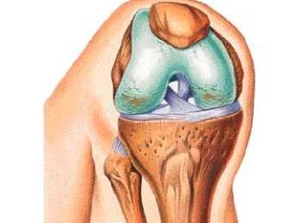
Osteoarthrosis or osteoarthrosis of the knee joint is a disease that occurs on the basis of dystrophic changes with the subsequent growth of connective tissue.There are many factors that affect the development of the disease, but all of them ultimately lead to a breach of metabolism in the cartilage.In the medical literature, arthrosis of the knee joint is called gonarthrosis.
According to statistics, gonarthrosis occupies a leading position in the frequency of occurrence among other arthrosis.The disease brings severe discomfort that can develop into pain when walking and at rest.
Knowledge of early symptoms will help suspect the development of pathology and cure it in the initial stages.
Causes
According to medical classification, there is primary and secondary gonarthrosis or arthrosis of the knee joint.
Osteoarthritis of the knee joint can occur on the basis of various diseases or act as their complication.When the exact cause is not established due to an unclear history or clinical image, gonarthrosis is called primary, but if the cause is known, such arthrosis is called secondary.
Osteoarthritis develops with age in almost all people, on average this period begins after 45-50 years of life.
The course and pathogenesis of primary and secondary arthosis are the same and does not depend on the cause of the occurrence.
The most common causes of arthrosis and osteoarthrosis in the knee joint are:
- traumatic injury to the knee;
- Common deformation inward and outside;
- abbreviation of an lower extremity;
- abnormal hypermobility in the joint;
- chondroblast dysplasia;
- Calcinosis of cartilage;
- Osteomyelitis of the femur and tibia;
- rheumatoid arthritis or arthritis of any other etiology;
- glucose metabolism;
- Metabolic diseases and hormonal diseases.
Damage.After receiving the knee injury in the joint cavity, inflammation with a strong focus on alitation can develop.After the disappearance of pro -inflammatory agents, the processes are activated with replacement or arthrosis.
Most often, the disease occurs on the basis of a fracture with a rupture of the ligament and damage to the bag and the surface of the articular cartilage.
Congenital deformations.Valgus or Varior deformation is found very often, and without proper correction it can be complicated by sclerotic changes in the knee.This happens due to the fact that one of the knees falls more than the load than it should be.
Abbreviation of any of the lower extremities.In addition to deformations, in the pathogenesis of the development of the disease, incorrect distribution of weight on the knees plays the role.
Hypermores of the knee.In this condition, wear of the cartilage tissue in the knee joint can occur with subsequent degeneration and degeneration in arthrosis.Hypermorsion often leads to spontaneous dislocations and sprains of the joint capsule.
Hand -haired dysplasia.Due to the wrong development of the motor surface of the knee joint, a pathological growth of connective tissue occurs.
Calcinosis of the joint.The pathogenesis is based on deposition of salts in the joint and the formation of a specific precipitate, causing calcification with subsequent osteoarthritis.
Osteomyelitis.Inflammatory bacterial disease where the destruction of bones and joints occurs.First, ankylosis is formed and only then sclerosis.
Arthritis of any etiology.The most dangerous are rheumatoid arthritis, accompanied by autoimmune lesions in the heart and joint.
Diabetes, such as metabolic disorders, leads to a violation of the flow of nutrients in the joint and spire of cartilage.
Obesity.With a great weight of the body, large loads are on the knees as they walk and in a standing position.As a result of constant pressure, the blood flow drops to the knee joints and atrophy with dystrophy develops.
Symptoms
Symptoms of arthrosis of the knee joint depend on the stage of the pathological process.Based on this, analysis of the symptoms and pace of their growth, you can assess the extent of changes in cartilage tissue.
Symptoms of knee osteoarthritis:
- the presence of pathological sounds during movement;
- tenderness after load or at rest;
- Reduction of features;
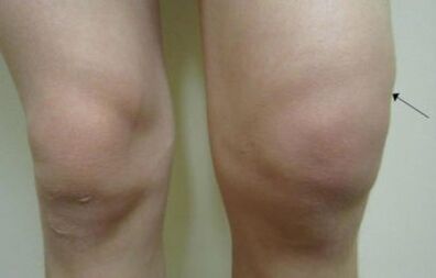
- edema and common increase;
- pathological dislocations, fractures and subluxation;
- Temporary jamming that may occur when flexion and expansion of the joint.
Clicks and crunches are not noticed immediately and if they notice, they do not add duly attention.Pathological sounds encounter the idea that a pathological process of deposition of salts or the formation of osteophytes occurs in the cartilage gap.
Pain occurs due to the formation of calcinates or osteophytes.First, the pain syndrome is not expressed, later it only appears in the morning and passes after lunch, with the development of the disease, pain may occur at rest.
A decrease in joint functions is manifested in the stiffness of movements and a decrease in their amplitude.Depending on the scene, the restriction of movements may last a specific time and pass at rest.
Edema occurs due to inflammation and hypersecretion of the synovial fluid.There are also options when the skin is inflamed over the joints.Such symptoms can be with scarlet fever or arthritis.
Dislocations and subluxation occur for the reason that the process applies to bones and ligaments on the knees.
Clushing is a condition where the movement in any axis is completely limited.Such a symptom indicates the neglect of the process and the need for complex treatment.
Degree of arthrosis
Classify osteoarthritis according to the following signs:
- radiological symptoms;
- clinical manifestations;
- Laboratory data.
The most common and practical classification is radiological, it is simple and understandable, even for people without medical education.
Based on x -rays four degrees of arthosis in the knee joint is separated:
- Reduction of the common hole is small and there are no osteophytes;
- The interpoint gap is not tied, but there are signs of small calcinates or osteophytes;
- The Inspoint -Gorge has a narrowing expression that is osteophytes, the joint formation begins;
- Lack of common gap, bone formation, ankylosis and dystrophy.
With regard to the clinical image, the following phases are separated:
- Mild degree symptoms are insignificant, occur in the morning and pass 30-60 minutes after waking up;
- The average degree is a significant symptom, the feeling of discomfort goes before lunch, the swelling is insignificant, it quickly steps out without treatment;
- A serious degree - characterized by constant sore pain, discomfort at rest, morning stiffness does not go until noon, ankylosis, burgely and sinusitis in the knee joint develops.
Laboratory studies are taken into account, the indicators of soukocytes are evaluated.It is also necessary to check the presence of a rheumatoid factor.
Diagnostic methods
The diagnosis of arthrosis of the knee joint is not complicated but requires certain skills from the doctor.
Two types of diagnostic measures are separated:
- Laboratory diagnostics;
- Instrumental diagnostics.
For the correct diagnosis, each of the methods must be taken into account and analyze the image as a whole.
Laboratory
If osteoarthritis is suspected, the participating physician prescribes the following tests:
- General blood and urine test;
- biochemical blood test;
- Determination of antibodies to the rheumatoid factor;
- Determination of antibodies to their own cells.
Laboratory data does not perform information about the development stage of the disease.
Starring
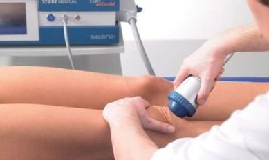
The instrumental diagnosis of arthrosis includes the following methods:
- Radiography in two standard projections;
- minimally invasive arthroscopy;
- UZD study;
- Ct;
- Mr;
- Scintigraphy (according to indications).
Radiation diagnosis is aimed at determining changes in the joint and to assessing the condition of the cartilage.
Treatment
Treatment of osteoarthritis of the knee joint is a long process.The duration of treatment is due to the fact that the replacement of cartilage tissue occurs very slowly, and in some cases it is completely impossible to restore the joint.
Modern methods of treating arthrosis in the knee joint include extensive goals aimed at removing inflammation, normalizing the lifestyle and improving metabolism in cartilage tissue.
There are such therapy methods:
- Drug therapy;
- Exercise therapy and massage;
- folk medicine;
- Surgical intervention.
The doctor prescribes treatment based on the duration of the disease, the stage of its development and clinical manifestations.
Medicines
Drug therapy is aimed at relieving pain and inflammatory reaction.For this purpose, the following drugs are prescribed:
- Non -steroidal anti -inflammatory;
- Chondroprotectors;
- glucocorticoid;
- Cytostatic.
Tablets from arthrosis of the knee joint have many side effects, in the treatment it is necessary to monitor the condition of the stomach and the kidneys.
Often, medicine for arthrosis is prescribed for a long period of time, so the least toxic drugs must be chosen.
Exercises
Treatment of arthosis using exercise therapy aims to strengthen the muscles and ligament of the knee.With dosed loads in the cartilage of the diseased joint, metabolism improves and regenerative processes are accelerated.
Exercises must be selected individually taking into account the stage of the patient's illness and physical abilities.
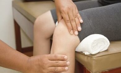
Massage
Massage of the knee joint allows you to improve blood flow and relieve discomfort.Proper massage can prevent the appearance of ankyloses and false joints.
People
Treatment of arthrosis of the knee joint at home should not be the most important method of combating pathology or can only act as an addition to the drug.
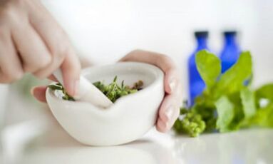
House processing includes:
- decrease in body weight;
- Normalization of working hygiene and compliance with today's regime;
- The fight against inflammation.
The anti -inflammatory effect is occupied by such herbs:
- Tincture from thyme and St.John's Wort;
- burdock leaves;
- White cabbage leaves;
- Infusions and decoction from dandelion and chamomile.
Operation
The operation is prescribed with the inefficiency of conservative therapy or at the request of the patient.One of the most important indications of surgical intervention is the 4th phase of the disease of radiological properties.
During surgery, surgeons can completely replace the joint with an endoprotesis or change one of its parts.